A gastric polyp is a visible growth projecting from the stomach lining, but that description does not identify its cause or cancer risk. By the end, you will know how doctors classify these lesions, which symptoms and endoscopic findings demand closer assessment, and why biopsy results—not appearance alone—determine whether observation or removal is appropriate.
Key takeaways
- A gastric polyp is a growth seen above the stomach lining, not a diagnosis.
- Fundic-gland polyps, hyperplastic polyps and adenomas carry different cancer risks.
- Endoscopy assesses size, location and surface features before biopsy or removal.
- Pathology determines whether you need surveillance, treatment or no further follow-up.
What a Gastric Polyp Is—and What It Is Not
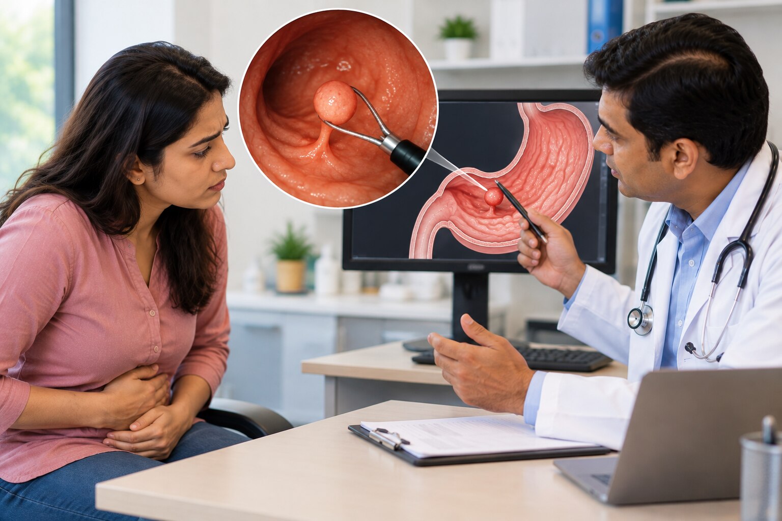
A gastric polyp is an endoscopic description, not one disease. It means tissue projecting above the stomach lining, but the growth may be a fundic-gland polyp, hyperplastic polyp, adenoma, inflammatory lesion, or another condition. Their causes, cancer risk, and need for removal differ.
The phrase “gastritis versus gastric polyp body” captures a common confusion: gastritis affects the stomach lining broadly, while a polyp is a discrete raised growth.
| Finding | What it means | Key distinction |
|---|---|---|
| Stomach polyp | A raised growth from the stomach lining | Describes appearance, not a final diagnosis |
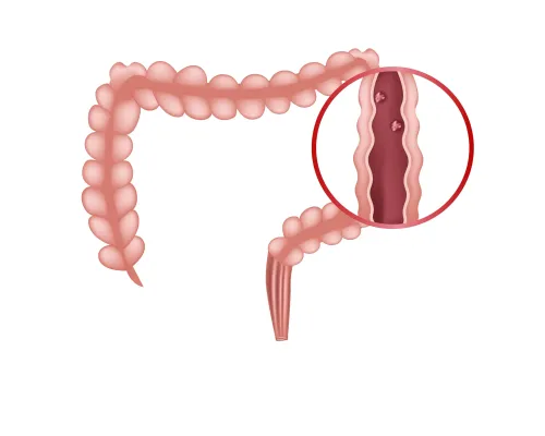
| Colorectal polyp | A growth in the colon or rectum | Different organ, tissue context, and cancer-risk assessment |
| Gastritis | Inflammation of the stomach lining | Not a discrete mass, although it can surround a polyp |
| Peptic ulcer | An open break in the stomach or duodenal lining | A crater-like defect rather than a projecting growth |
| Stomach cancer lesion | Malignant tissue in the stomach | May appear irregular, ulcerated, thickened, or polyp-like and requires biopsy assessment |
Small polyps often cause no symptoms because they do not disrupt digestion or bleed. Do not assume a finding is harmless if you have:
- Upper abdominal pain
- Nausea or vomiting
- Unexplained iron-deficiency anaemia
- Black, tarry stools
- Unintentional weight loss
These symptoms need medical assessment because bleeding, obstruction, inflammation, or cancer can produce them. Symptom-free status alone does not identify the polyp’s type.
The Main Types of Gastric Polyp and Their Cancer Risk
A solitary, typical fundic gland polyp has very low malignant potential, but the risk assessment changes with its pattern, pathology, and the stomach around it.
| Type | Typical location | Common associations | Cancer risk |
|---|---|---|---|
| Fundic gland polyp | Body or fundus | Proton-pump inhibitor use; familial adenomatous polyposis when numerous or found in a young person | Very low for a typical sporadic lesion; dysplasia, numerous lesions, or an atypical appearance needs further evaluation |
| Hyperplastic gastric polyp | Any area, often where the lining is inflamed | H. pylori, chronic gastritis, autoimmune gastritis, atrophy, and intestinal metaplasia | Usually low, but dysplasia and surrounding mucosal changes increase concern |
| Adenomatous polyp | Often the antrum, though it can occur elsewhere | Atrophic gastritis and intestinal metaplasia | Premalignant; complete removal is generally required |
Eradication of H. pylori can sometimes shrink or eliminate an H. pylori gastric polyp. Biopsies from normal-looking stomach lining matter because atrophy or intestinal metaplasia can raise long-term cancer risk even when the polyp shows no dysplasia.
Use these findings to decide whether a common-looking lesion needs a wider evaluation:
- Numerous fundic gland polyps
- A young patient with multiple lesions
- A personal or family history of familial adenomatous polyposis
- Dysplasia, ulceration, bleeding, large size, or an irregular surface
Gastric neuroendocrine tumors can resemble polyps but need separate management. Type 1 and type 2 tumors may be multifocal and linked to hypergastrinemia; type 3 tumors are usually sporadic and carry greater invasion or metastasis risk. Treatment depends on type, size, number, depth, and evidence of spread.
How Upper Endoscopy Finds and Investigates a Polyp
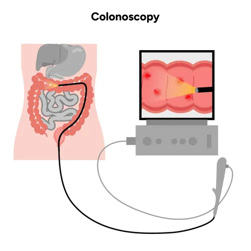
Upper GI endoscopy examines the oesophagus, stomach, and duodenum while the endoscopist records each lesion’s size, number, location, colour, surface, attachment, ulceration, and surrounding mucosa. A gastroscopy gastric polyp report should capture these details because they determine whether sampling or removal is appropriate.
Appearance may suggest a diagnosis but cannot prove it. The same visual finding can represent a polyp, ulcer, inflamed tissue, or a malignant gastric lesion.
| Appearance | What it may suggest | Why it is not proof |
|---|---|---|
| Smooth, pale, elevated lesion | Fundic gland tissue or another polyp | A small cancer focus can remain hidden |
| Red, eroded, or ulcerated lesion | Inflammation, hyperplastic change, or cancer | Ulceration increases concern but does not identify the cause |
| Irregular or broad-based lesion | Dysplasia, adenoma, or invasive disease | Depth and cell changes require tissue assessment |
A gastric polyp biopsy uses forceps to collect tissue even when immediate removal is not planned. Pathology can identify hyperplasia, inflammation, dysplasia, adenoma, fundic gland tissue, or cancerous change, then guide surveillance or the next procedure.
A small sample is not always representative. It can miss focal dysplasia or carcinoma in a large, irregular, ulcerated, or broad-based lesion. When the lesion can be removed safely, complete excision provides a more definitive diagnosis because the entire surface and deeper tissue can be examined.
When Observation Is Reasonable—and When Removal Is Preferred
A typical, small lesion with reassuring histology, no symptoms, and normal surrounding mucosa may suit gastric polyp observation with planned reassessment. The decision also weighs size, number, location, attachment, surface appearance, depth, bleeding, background atrophy or intestinal metaplasia, and personal risks such as hereditary polyposis.
Complete gastric polyp removal is preferred for:
- Adenomas or any dysplasia
- Persistent or symptomatic hyperplastic polyps, especially those around 1–2 cm or larger
- Bleeding, ulcerated, irregular, broad-based, or rapidly changing lesions
- Polyps with an unreliable or limited biopsy, because forceps samples can miss focal cancer
- Lesions in a high-risk stomach with atrophy or intestinal metaplasia
Technique depends on the lesion, not size alone.
| Option | What it means | When it applies |
|---|---|---|
| Snare removal | Cuts a stalked or suitable superficial polyp, sometimes after lifting it | Small, accessible lesions that can be removed completely |
| Endoscopic mucosal resection | Lifts and removes superficial tissue, often in one piece or pieces | Selected superficial neoplasia without suspected deep invasion |
| Endoscopic submucosal dissection | Dissects beneath the lesion for en-bloc removal | Larger or flat superficial lesions where precise histology matters |
| Surgery | Removes part or all of the stomach | Suspected deep invasion, extensive disease, or unsuitable anatomy |
Anticoagulants, antiplatelet drugs, bleeding disorders, and major comorbidities alter timing and preparation. They do not prove what the polyp is; the treating clinician must balance bleeding from removal against harm from stopping medication.
What the Pathology Report Changes About Your Follow-Up
The pathology result turns an uncertain visual finding into a tailored gastric polyp follow-up plan. “No dysplasia” is reassuring, but it does not end evaluation if the lesion was incompletely removed or the surrounding stomach shows high-risk changes.
| Finding | What it means | What it may change |
|---|---|---|
| Hyperplastic change or inflammation | Often reflects mucosal injury, H. pylori, or gastritis | Test and treat H. pylori; remove a persistent, large, bleeding, or suspicious lesion |
| Fundic gland polyp | Usually low risk when solitary and typical | Observation may be enough; atypical, large, numerous, ulcerated, symptomatic, or dysplastic lesions need further assessment |
| Adenoma or gastric dysplasia | A premalignant finding that sampling can underestimate | Complete endoscopic removal, review of the remaining stomach, and repeat endoscopy |
| Cancerous change | Malignant tissue or possible invasion | Additional sampling, staging scans, and specialist treatment planning |
Biopsies from apparently normal mucosa can reveal atrophic gastritis or gastric intestinal metaplasia. Those findings create a separate cancer-risk question, even when the polyp itself is benign; gastric intestinal metaplasia may justify surveillance based on its extent, family history, and other risks.
A benign solitary lesion with healthy surrounding mucosa does not automatically require repeated procedures. Hereditary polyposis, incomplete removal, gastric dysplasia, persistent bleeding, a suspicious appearance, or extensive atrophy requires closer planning. With Dr Pravish Ghurde Gastroenterologist in Mulund, ask which diagnosis, procedure, and follow-up match both the pathology and your overall gastric risk.
Frequently asked questions
What is a gastric polyp?
A gastric polyp is tissue projecting above the stomach lining seen during upper endoscopy. It can represent several different conditions.
Which gastric polyps have the highest cancer risk?
Adenomas carry the greatest cancer concern among the common types. Hyperplastic and fundic-gland polyps have different risks that depend on their features and cause.
How does upper endoscopy investigate a gastric polyp?
Upper endoscopy shows the polyp’s size, location and surface. The endoscopist can take a biopsy or remove the lesion for laboratory examination.
When is gastric polyp removal preferred?
Removal is preferred when the polyp has concerning features, is an adenoma, or requires complete examination. Small lesions without concerning features may be observed.