Gluten can trigger an immune attack that damages the small-intestinal lining, reducing the surface available to digest food and move nutrients into the bloodstream. By the end, you will understand which deficiencies to look for, how celiac disease differs from other gluten-related conditions, how testing works, and what doctors investigate when malabsorption continues.
Key takeaways
- Celiac disease damages intestinal villi through an immune response to gluten.
- Check iron, folate, vitamin B12, calcium, and vitamin D after diagnosis.
- Keep eating gluten before testing unless your clinician tells you to stop.
- Persistent malabsorption requires assessment for ongoing exposure or another digestive disorder.
How gluten damages the small intestine
Gluten does not simply irritate the gut in celiac disease; it starts an immune attack against the lining of the small intestine. During digestion, gluten releases peptides that cross the intestinal barrier. In genetically susceptible people, immune cells recognise these peptides as dangerous and activate a gluten-triggered immune response.
The response involves T cells, inflammatory signals and antibodies directed at tissue transglutaminase. It also increases intraepithelial lymphocytes, immune cells positioned between the lining’s surface cells. These cells injure the epithelium, while inflammation disrupts the normal renewal of the intestinal lining.
The characteristic damage is villous atrophy: the finger-like villi become shortened, flattened or lost. At the same time, the glands between villi enlarge and produce excess immature cells, a change called crypt hyperplasia. The surface becomes less efficient at moving digested nutrients from the intestine into the bloodstream.
That loss of absorptive area turns a microscopic injury into malabsorption. Enzymes attached to the intestinal surface can work less effectively, and damaged cells cannot transport nutrients normally. You might have diarrhoea, bloating or abdominal pain, but digestive symptoms are not required; impaired absorption can first appear as unexplained anaemia, weight loss or fatigue.
Continued gluten exposure keeps the immune process active, so the injury can persist even when each meal causes little noticeable discomfort.
Which nutrients and body functions are affected
Villous atrophy removes absorptive surface, while crypt hyperplasia replaces mature transport cells with less effective ones. The result is malabsorption: nutrients cross the small-intestinal lining less efficiently, even when digestion itself seems normal.
| Nutrient | What poor absorption can cause | Physical finding |
|---|---|---|
| Iron | Reduced haemoglobin production | Iron-deficiency anaemia, fatigue, mouth ulcers |
| Folate | Impaired red-cell production and cell division | Anaemia, mouth ulcers |
| Vitamin B12 | Nerve and blood-cell problems; absorption mainly occurs in the terminal ileum | Neuropathy, anaemia |
| Calcium and vitamin D | Poor mineral supply and weakened bone formation | Muscle weakness, bone loss |
| Magnesium and zinc | Impaired muscle, nerve, immune and tissue functions | Muscle weakness, poor healing |
| Vitamins A, E and K | Reduced vision and nerve protection, antioxidant activity and clotting | Neuropathy, bruising |
Weight loss reflects both nutrient loss and reduced usable energy. Vitamin B12 deficiency is possible, but damaged duodenal villi alone do not explain it; persistent low B12 needs assessment of terminal-ileum absorption and other causes.
Lactase sits on the intestinal brush border, so villous injury can produce temporary lactose intolerance with bloating or diarrhoea. It often improves as the lining heals, so permanent dairy avoidance is not automatic; if dairy is restricted, protect calcium and vitamin D intake. Correcting deficiencies does not stop ongoing intestinal injury from gluten exposure.
How celiac disease differs from other gluten reactions—and how testing works
Celiac disease is an autoimmune disorder; non-celiac gluten sensitivity causes symptoms without the characteristic intestinal injury or antibody pattern, while wheat allergy is an IgE-mediated reaction to wheat proteins. The timing and test results separate them.
1. Keep gluten before testing. A gluten-free diet can lower antibody levels and partly heal the small intestine, producing misleading results. Discuss testing with a clinician before removing gluten.
2. Start with tissue transglutaminase IgA (tTG-IgA) and total IgA. If IgA deficiency makes the IgA result unreliable, ask about deamidated gliadin peptide IgG or tTG-IgG. Endomysial antibody provides additional support. A negative tTG-IgA does not fully exclude celiac disease when gluten exposure is limited, disease is mild or patchy, or suspicion remains high.
3. Confirm suspected celiac disease with an upper endoscopy and duodenal biopsy when indicated. A wheat allergy is investigated with an allergist’s history, skin-prick testing or wheat-specific IgE; hives, wheezing, throat tightness or vomiting soon after wheat points toward allergy.
Non-celiac gluten sensitivity has no validated blood test and is diagnosed only after celiac disease and wheat allergy are excluded, followed by symptom assessment during structured dietary changes.
Pure, uncontaminated oats are tolerated by most people with celiac disease, but ordinary oats can carry wheat, barley or rye contamination. Choose gluten-free-labeled oats and report recurring symptoms or abnormal antibodies.
Restoring absorption after diagnosis
Once celiac disease is confirmed, the treatment that protects the intestine is a strict lifelong gluten-free diet. Do not merely reduce bread or avoid gluten on most days: even small exposures can restart immune injury, delay intestinal healing and keep nutrient absorption poor.
Avoid wheat barley and rye, including ingredients derived from them. Check gluten-free-labeled foods, ask restaurants about preparation, and prevent cross-contact from shared toasters, chopping boards, sieves, frying oil and food containers.
Oats require a separate decision. Pure uncontaminated oats are tolerated by most people with celiac disease, but ordinary commercial oats can contain wheat, barley or rye. Choose gluten-free-labeled oats and report renewed symptoms; follow-up antibody testing can help assess whether they are suitable for you.
- Replace documented deficiencies with clinician-directed nutrient replacement, such as iron, folate, vitamin B12, calcium or vitamin D. Avoid taking high-dose supplements indefinitely without blood-test guidance.
- Review recovery with symptoms, weight, blood counts, iron studies and celiac antibodies. Symptoms can improve within days or weeks, while intestinal healing often takes months or longer in adults.
- If problems continue, investigate ongoing gluten exposure, lactose intolerance, irritable bowel syndrome, microscopic colitis, pancreatic disease or an incorrect diagnosis before adopting a more restrictive diet. Refractory celiac disease is uncommon and requires specialist assessment, not self-diagnosis.
When ongoing malabsorption needs a wider investigation
Persistent symptoms do not automatically mean you need a stricter gluten-free diet. Doctors first check for continuing gluten exposure, an incorrect original diagnosis, or another digestive disorder before considering rare refractory celiac disease.
A wider review may include:
- A diet review with a trained clinician, including sauces, supplements, restaurant meals, shared toasters, and cross-contact.
- A complete blood count, iron studies, folate and vitamin B12 levels, calcium, vitamin D, magnesium, zinc, and liver tests to identify untreated or recurring deficiencies.
- Stool tests for infection, inflammation, blood, or excess fat when diarrhoea, weight loss, or greasy stools continue.
- Testing for lactose intolerance or other carbohydrate intolerance when bloating and diarrhoea follow specific foods.
- Assessment for irritable bowel syndrome when symptoms persist without evidence of intestinal inflammation.
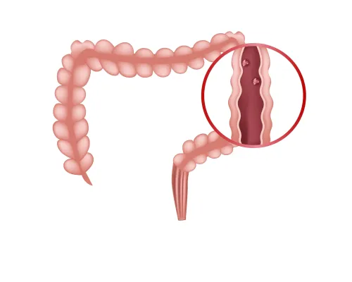
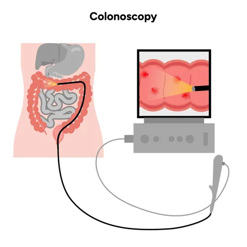
- Colon evaluation for microscopic colitis, particularly with chronic watery diarrhoea.
- A pancreatic evaluation when stool fat, weight loss, or upper-abdominal symptoms suggest pancreatic disease.
- Review of medicines, thyroid function, inflammatory bowel disease, and other causes of malabsorption when initial results do not explain the problem.
Iron-deficiency anaemia deserves attention even without diarrhoea, especially when oral iron fails or anaemia returns. Dr Pravish Ghurde Gastroenterologist can help coordinate testing when persistent malabsorption overlaps with inflammatory bowel or pancreatic disease.
Refractory celiac disease is uncommon and requires specialist confirmation; do not diagnose it from symptoms alone. Correcting low nutrient levels also does not replace strict gluten avoidance if exposure continues to injure the intestine.
Related service
Malabsorption Syndrome Treatment Malabsorption Syndrome Treatment Dr. Pravish Ghurde offers comprehensive diagnosis and effective treatment for malabsorption syndrome at Platinum Hospital, Mulund, Mumbai. View service → |
Frequently asked questions
How does gluten damage the small intestine in celiac disease?
Gluten triggers an immune response that injures the small-intestinal lining and flattens villi, reducing the surface available for digestion and absorption.
Which nutrients can celiac disease affect?
Damage can reduce absorption of iron, folate, vitamin B12, calcium, vitamin D, and other nutrients, contributing to anaemia, bone loss, fatigue, or nerve symptoms.
How is celiac disease different from other gluten reactions?
Celiac disease is an autoimmune disorder with intestinal injury. Wheat allergy involves an allergic response, while non-celiac gluten sensitivity does not produce the same confirmed intestinal damage.
How is celiac disease tested?
Testing usually starts with tissue transglutaminase IgA and total IgA blood tests while you are eating gluten; a specialist may confirm the diagnosis with a small-intestinal biopsy.
What helps restore nutrient absorption after diagnosis?
A strict, lifelong gluten-free diet allows the intestinal lining to heal. Follow-up blood tests and replacement of documented deficiencies help track recovery.