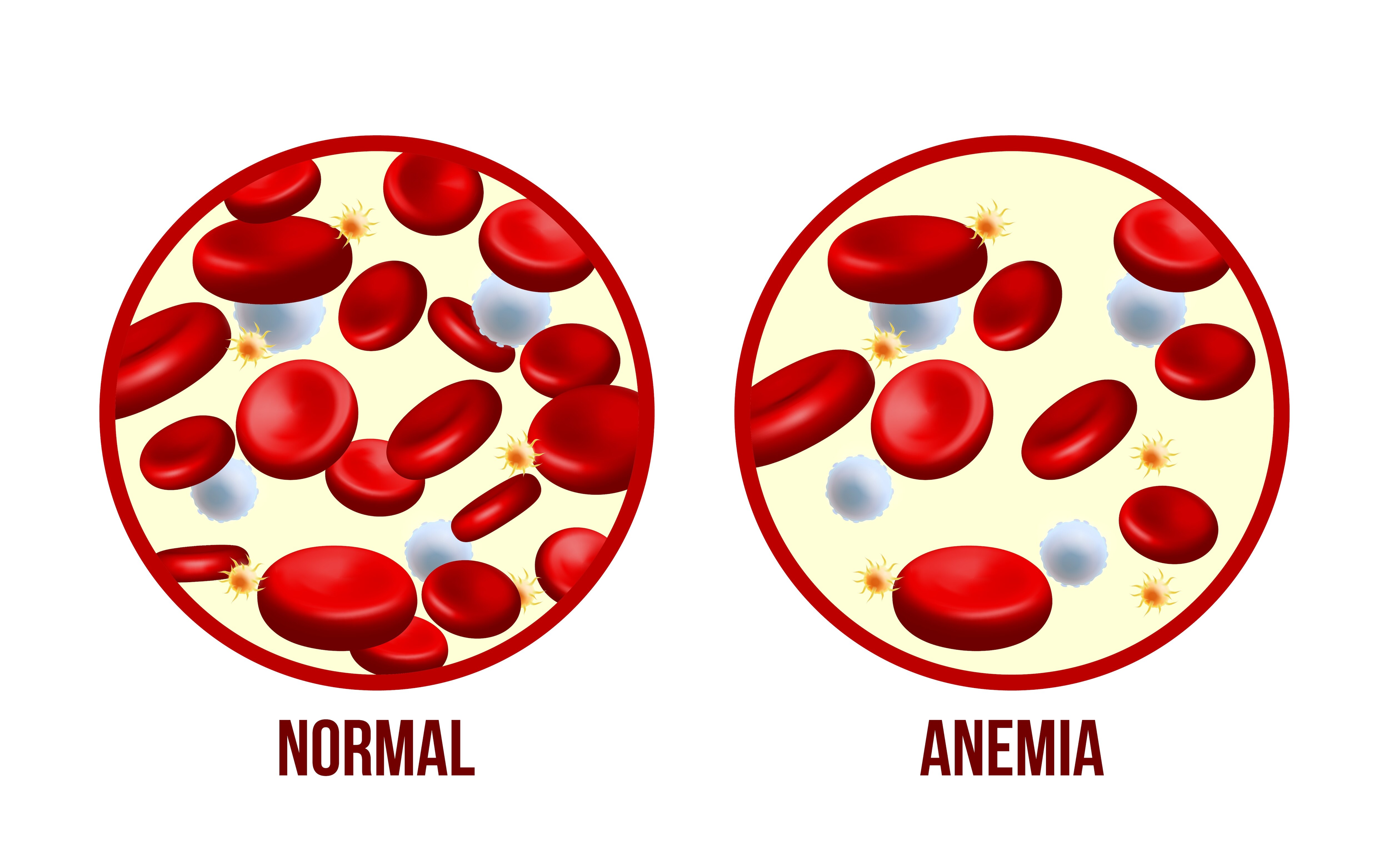
Fatigue alone cannot show whether you are short of iron, losing blood, or absorbing nutrients poorly. By the end, you will know which blood results confirm iron deficiency, how gastrointestinal disease causes microscopic blood loss, and when endoscopy or further testing deserves prompt attention.
Key takeaways
- Confirm iron deficiency with ferritin and transferrin saturation, not MCV alone.
- Digestive disease can cause microscopic blood loss or prevent iron absorption.
- Recurrent anemia needs investigation even when you see no blood in stool.
- Negative endoscopies may require medication review, small-bowel testing, or repeat evaluation.
How blood tests confirm iron deficiency anemia
A complete blood count confirms anemia by showing low haemoglobin; a low mean corpuscular volume (MCV) supports iron restriction but does not prove it.
Confirm the cause with iron studies: ferritin below 45 ng/mL (45 µg/L) in an anemic patient strongly supports iron-deficiency anemia (IDA), especially when transferrin saturation is below 20% and total iron-binding capacity is raised.
| Pattern | Ferritin | Transferrin saturation and TIBC | Other clues |
|---|---|---|---|
| Iron-deficiency anemia | Below 45 ng/mL | Saturation low; TIBC high | Low haemoglobin; MCV often low; red cells may be pale |
| Inflammation-related anemia | Normal or high | Saturation low; TIBC low or normal | C-reactive protein may be raised; ferritin can mask depleted stores |
| Vitamin B12 or folate deficiency | Normal or high | Usually not the defining finding | MCV high; numbness or a sore tongue can occur |
| Kidney-disease anemia | Usually normal | Variable | Low kidney function; MCV often normal |
| Thalassemia trait | Normal | Usually normal | Very low MCV with a relatively preserved red-cell count; lifelong pattern |
Serum iron changes with meals, timing and illness, so interpret it with ferritin, transferrin saturation and TIBC rather than alone. Infection or inflammation can make ferritin appear normal or high; C-reactive protein and a low transferrin saturation help reveal iron deficiency hidden by that response. A normal MCV also does not exclude early deficiency.
How gastrointestinal disease causes iron loss without visible bleeding
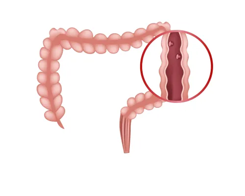
Normal-looking stool does not rule out occult gastrointestinal bleeding. A lesion can release only microscopic blood loss into the digestive tract, diluted through stool or occurring intermittently, so you never see red blood or black, tarry stools. Over weeks or months, that loss can deplete iron stores faster than your diet replaces them.
Common sources include:
- Ulcers in the stomach or duodenum, which can ooze slowly rather than bleed heavily.
- Medication-related erosive gastritis or small-bowel injury from ibuprofen, naproxen, aspirin, or combination pain and cold medicines.
- Colorectal polyps and cancer, which can bleed intermittently.
- Inflammatory bowel disease, where inflamed bowel lining loses blood and iron.
- Vascular lesions, which can produce small, recurring leaks.
Iron deficiency can also develop without blood loss. Reduced stomach acid can impair the chemical steps that release iron from food, while celiac disease damages the small-intestinal lining where iron is absorbed. Other small-intestinal disorders can have the same effect, even without diarrhoea or weight loss.
Helicobacter pylori infection can contribute through chronic gastritis, impaired iron handling, and ulcers. Because bleeding and absorption problems can coexist, a negative stool blood test does not by itself exclude a gastrointestinal cause.
Which symptoms and risk factors make a gastrointestinal source more likely
Persistent fatigue, exertional breathlessness, palpitations, or pica (ice or non-food cravings) deserve medical review when iron deficiency is confirmed. Gastrointestinal evaluation becomes more important with:
- unintentional weight loss, altered bowel habits, persistent abdominal pain, or recurrent indigestion
- vomiting blood, black tarry stools, or maroon or bright-red rectal bleeding
- regular ibuprofen, naproxen, aspirin, or combination pain and cold medicines
- a family history of bowel or stomach cancer, or anemia that returns after iron treatment
| Clue | Why it matters | How quickly to seek evaluation |
|---|---|---|
| Men and postmenopausal women | There is no menstrual explanation for ongoing iron loss | Arrange gastrointestinal assessment promptly |
| Menstrual blood loss, pregnancy, diet, or blood donation | These can reduce iron stores, but do not rule out an ulcer, bowel lesion, or absorption problem | Discuss the full history; persistent or recurrent anemia needs further investigation |
| Vomiting blood, black tarry stools, maroon or bright-red rectal bleeding, fainting, chest pain, severe breathlessness, or marked weakness | These can indicate active bleeding or dangerously reduced oxygen delivery | Seek emergency care now |
Do not assume that improved blood counts prove diet or menstrual blood loss was the only cause. Iron can temporarily correct anemia while an ulcer, cancer, inflammatory lesion, or malabsorption disorder continues.
Premenopausal women with persistent anemia also need an individualized discussion of gastrointestinal testing, based on symptoms, age, menstrual blood loss, family history, and anemia severity.
What gastrointestinal evaluation usually involves
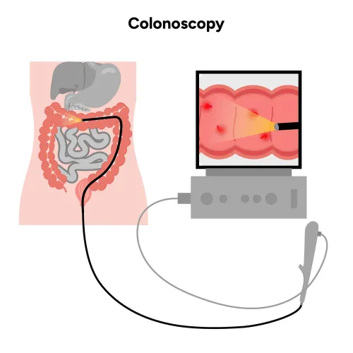
After iron-deficiency anemia is confirmed, the usual search examines the stomach, small intestine and colon. In adult men and postmenopausal women, this usually means bidirectional endoscopy: colonoscopy and upper endoscopy, often during the same session, even without pain, visible blood or altered bowel habits.
| Examination or test | What it can identify | When it applies |
|---|---|---|
| Colonoscopy | Polyps, colorectal cancer, ulcers and inflammatory bowel disease | Usually part of initial evaluation |
| Upper endoscopy | Esophagitis, gastritis, ulcers and upper-gastrointestinal tumors | Usually paired with colonoscopy |
| Celiac tissue-transglutaminase IgA and total IgA | Celiac-related malabsorption | Test while eating gluten; avoiding gluten can cause a misleading negative result |
| Helicobacter pylori noninvasive testing | Infection linked to gastritis, impaired iron handling and ulcer bleeding | After negative endoscopy, rather than routine stomach biopsies solely for H. pylori |
| Video capsule endoscopy | Bleeding, ulcers or inflammation in the small-bowel lining | When anemia persists, returns or shows ongoing blood loss after negative endoscopies |
| CT enterography or MR enterography | Small-bowel masses, narrowing and inflammatory disease | Complements capsule testing when structural disease is suspected |
A fecal immunochemical test (FIT) detects blood shed into stool, but a negative result does not rule out colorectal cancer and is not a replacement for endoscopic evaluation. Its limitation includes intermittent bleeding and lesions outside the colon.
Menstruation can explain iron loss, yet persistent or severe anemia still warrants shared decision-making about gastrointestinal testing in premenopausal women.
What to do when anemia returns or the first tests are negative
An iron treatment response confirms that iron was limiting red-cell production; it does not prove that diet or menstrual loss was the only cause. Iron can raise haemoglobin while an ulcer, cancer, inflammatory lesion, or malabsorption disorder continues, so recurrent iron-deficiency anemia requires a renewed search rather than indefinite repeat supplementation.
If the first evaluation is negative, work through these checks with your clinician:
- Complete a medication review, including prescribed drugs, over-the-counter painkillers, and combination cold remedies. NSAIDs such as ibuprofen, naproxen and aspirin can cause stomach erosions, ulcers, and small-bowel injury with occult blood loss.
- Do not treat a negative fecal immunochemical test (FIT) as the end of the investigation. FIT can miss intermittent bleeding and does not assess upper-gastrointestinal or small-bowel disease.
- Test for celiac disease with tissue-transglutaminase IgA and total IgA while you are still eating gluten. Avoiding gluten before testing can produce a misleading negative result.
- Request renal-tract evaluation when no gastrointestinal source is found. Urinary blood loss and kidney or urinary-tract disease can accompany an unexplained anemia.
- Reconsider menstrual loss, family history, persistent symptoms, and whether upper and lower endoscopy were appropriate, including in premenopausal women.
- Ask a gastroenterologist whether small-bowel testing or repeat endoscopy is needed if anemia returns or iron stores fall again.
Dr Pravish Ghurde Gastroenterologist can help distinguish ongoing blood loss from malabsorption when routine results do not explain the pattern.
Related services
Malabsorption Syndrome Treatment Malabsorption Syndrome Treatment Dr. Pravish Ghurde offers comprehensive diagnosis and effective treatment for malabsorption syndrome at Platinum Hospital, Mulund, Mumbai. View service → |
Acute & Chronic Gastritis Dr. Pravish Ghurde provides expert diagnosis and comprehensive treatment for Gastritis at Platinum Hospital, Mulund, Mumbai. View service → |
Frequently asked questions
Which blood tests confirm iron deficiency anemia?
A complete blood count identifies anemia, while ferritin confirms depleted iron stores. Transferrin saturation, serum iron, total iron-binding capacity, and reticulocyte count help interpret the result, especially when inflammation affects ferritin.
How can gastrointestinal disease cause iron loss without visible bleeding?
Ulcers, gastritis, bowel polyps, cancers, inflammatory bowel disease, and vascular lesions can bleed in tiny amounts that mix with stool. Coeliac disease and other conditions can also reduce iron absorption without causing visible blood.
What symptoms and risk factors make a gastrointestinal source more likely?
Seek evaluation for recurrent anemia, black or bloody stools, abdominal pain, altered bowel habits, weight loss, persistent vomiting, swallowing difficulty, or unexplained fatigue. Risk also rises with older age, family history of bowel cancer, coeliac disease, inflammatory bowel disease, and regular NSAID use.
What does gastrointestinal evaluation usually involve?
Your clinician reviews medicines, diet, bowel symptoms, menstrual or other blood loss, and family history. Testing can include upper endoscopy, colonoscopy, coeliac blood tests, stool testing, and small-bowel imaging or capsule endoscopy when initial studies do not find a cause.
What happens when anemia returns or the first tests are negative?
Confirm recurrent iron deficiency, check treatment adherence and ongoing blood loss, and review medicines such as ibuprofen or aspirin. Your clinician may repeat endoscopy, test for coeliac disease or Helicobacter pylori, or investigate the small bowel.
Related services
Upper GI Bleeding Treatment Dr. Pravish Ghurde provides prompt and advanced care for upper gastrointestinal (GI) bleeding at Platinum Hospital, Mulund, Mumbai. View service → |
Crohn's Disease Dr. Pravish Ghurde offers expert diagnosis and comprehensive treatment for Crohn’s disease at Platinum Hospital, Mulund, Mumbai. View service → |
Colorectal Polyps, Lower GI Bleeding & Liver Disease Treatment Colorectal Polyps, Lower GI Bleeding & Liver Disease Treatment Dr. Pravish Ghurde, Consultant Gastroenterologist at Platinum Hospital, Mulund, Mumbai, provides expert diagnosis and advanced treatment for... View service → |