A stomach cancer can remain small and produce little more than recurring indigestion, yet the depth of invasion already determines which treatments are possible. By recognising warning signs, understanding personal risk, and knowing what endoscopy and biopsy establish, you can seek the right assessment before a treatable lesion becomes harder to cure.
Key takeaways
- Early gastric carcinoma is defined by invasion depth, not tumour size or symptom duration.
- Seek urgent care for vomiting blood, black stools, fainting or severe weakness.
- Endoscopy with biopsy confirms cancer; staging shows spread and guides treatment.
- Earlier diagnosis can expand treatment options and simplify follow-up planning.
What “early gastric carcinoma” means and why depth matters
Early gastric carcinoma is cancer confined to the mucosa or submucosa (T1), whether or not regional lymph nodes contain cancer. “Early” describes how deeply the tumour has invaded, not how long it has been present or how large it looks.
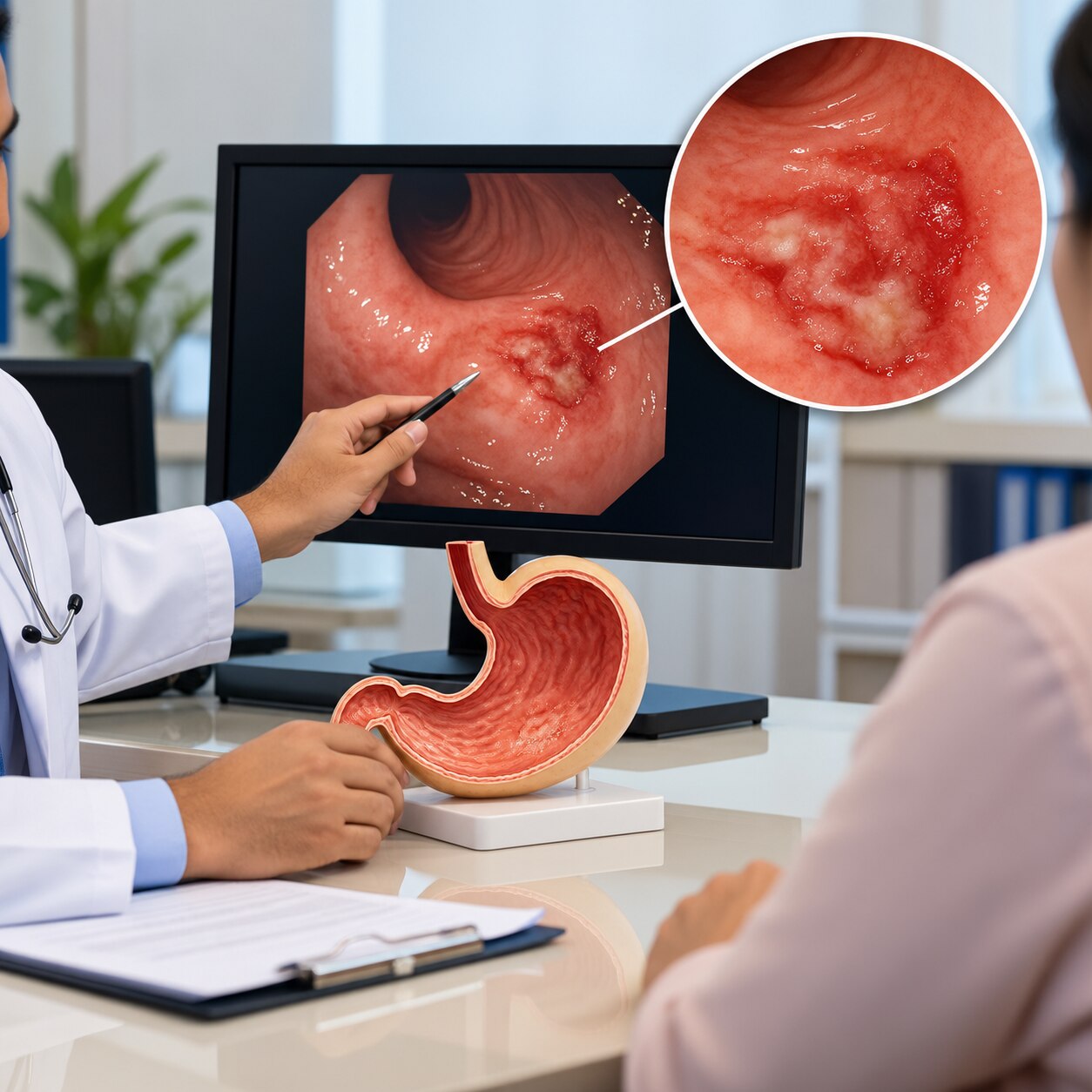
Early gastric cancer can appear as a flat, depressed or slightly discoloured area that is easy to miss.
Depth matters because deeper invasion increases the chance that cancer has reached lymphatic channels and nearby lymph nodes. A small tumour extending beyond the submucosa can therefore pose more danger than a wider lesion confined to the mucosa. Visible size still affects treatment decisions, but it cannot replace staging.
After endoscopic removal, the pathology report must establish whether the cancer is suitable for endoscopic treatment. Check that it records:
- The deepest layer invaded
- Whether the margins are clear
- How differentiated the cancer cells are
- Whether ulceration is present
- Whether lymphovascular invasion is present
A cancer limited to the mucosa, or meeting strict criteria within the submucosa (T1), can sometimes be cured with endoscopic mucosal resection or endoscopic submucosal dissection instead of stomach removal. Deep submucosal invasion, involved margins or other high-risk findings can require gastrectomy and lymph-node surgery, because the endoscopic specimen cannot treat cancer left elsewhere.
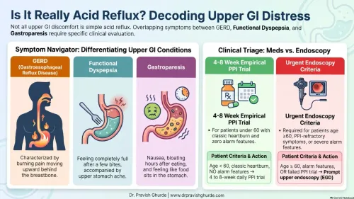
Which symptoms deserve assessment—and which require urgent care?
Gastric carcinoma may cause no symptoms at all, or only vague upper-abdominal discomfort, bloating, mild nausea or indigestion. Arrange assessment for persistent indigestion, early fullness after small meals, unexplained weight loss, recurrent vomiting, progressive difficulty swallowing, anaemia, or symptoms that keep returning. Feeling well between episodes does not rule out an early lesion.
These complaints can resemble gastritis, peptic ulcer disease or GERD because all can produce burning, nausea, bloating, pain, reflux or reduced appetite. Acid treatment may ease symptoms without explaining their cause, so improvement does not cancel an alarm symptom.
| Situation | What to do | Why it matters |
|---|---|---|
| Persistent or recurrent upper-abdominal symptoms, early fullness or weight loss | Book a prompt medical consultation | Symptoms need assessment even without severe pain |
| Progressive difficulty swallowing or suspected anaemia | Arrange medical assessment promptly | Ongoing bleeding or narrowing needs investigation |
| Vomiting blood, black tarry stools, fainting, severe weakness, severe abdominal pain, or vomiting that prevents fluids | Seek emergency care now | These signs can indicate gastrointestinal bleeding, dehydration or an acute complication |
Who needs a lower threshold for investigation?
Increasing age lowers the threshold for upper endoscopy, especially when indigestion, early fullness, anaemia, bleeding, vomiting, weight loss or swallowing difficulty persists. Do not wait for severe pain: early lesions can be subtle, and the absence of symptoms does not reliably exclude cancer.
Investigate sooner when you have:
- A family history of gastric cancer, particularly in a first-degree relative. A pathogenic CDH1 variant or a family pattern suggesting hereditary diffuse gastric cancer requires genetic counselling and specialist surveillance, not routine reassurance.
- Previous gastric biopsy findings such as atrophic gastritis, intestinal metaplasia, dysplasia or an incompletely characterised lesion.
- H. pylori infection, especially with atrophy or intestinal metaplasia. Eradication reduces future risk but does not replace endoscopy for alarm symptoms or a suspicious lesion.
- Pernicious anaemia, previous partial gastrectomy, heavy smoking, or origin from a region with high gastric-cancer incidence.
| Diagnostic evaluation | Population screening |
|---|---|
| Upper endoscopy is ordered because symptoms, anaemia, history or a finding creates individual concern. | Testing is offered to people without symptoms according to a country’s programme. |
| The examination directly inspects the stomach and biopsies suspicious areas. | Screening may use scheduled endoscopy in high-incidence settings such as Japan or South Korea. |
| A blood test, stool test or scan cannot replace biopsy of a suspicious lesion. | Routine screening is not recommended for every asymptomatic adult in countries where evidence is insufficient, including the United States. |
A normal examination lowers concern but does not erase risk when inherited or precancerous findings require planned follow-up.
How endoscopy, biopsy and staging establish the diagnosis
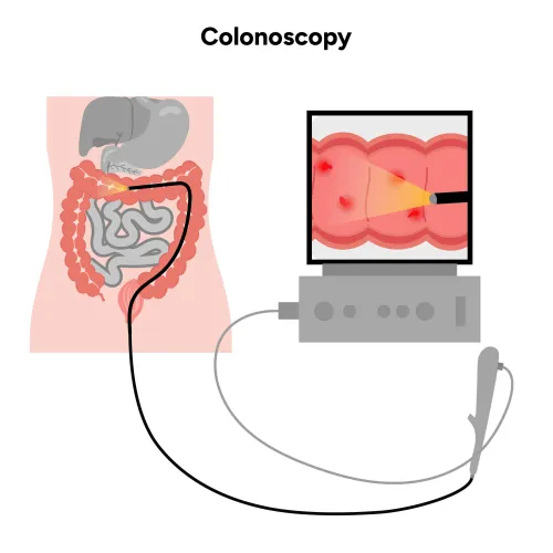
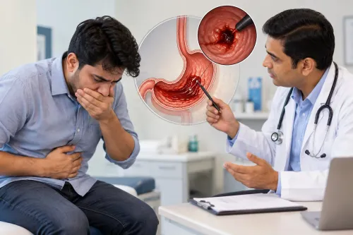
An upper endoscopy, also called esophagogastroduodenoscopy (EGD), allows direct inspection of the oesophagus, stomach and duodenum. It can reveal a flat, depressed or slightly discoloured lesion that scans miss. During the procedure, a targeted biopsy removes tissue from the suspicious area for microscopic examination.
A blood or stool test can suggest anaemia or bleeding, while a scan can show wall thickening or enlarged lymph nodes; neither can confirm malignant cells or identify the tumour’s cellular type.
A positive biopsy confirms gastric carcinoma, but it does not establish the complete stage. Doctors commonly use:
- Endoscopic ultrasound to measure invasion into the stomach wall and examine nearby lymph nodes; suspicious nodes can sometimes be sampled.
- Contrast-enhanced CT of the chest, abdomen and pelvis to assess regional lymph nodes and spread to organs such as the liver or lungs.
- PET-CT or staging laparoscopy when CT findings are unclear or doctors need to check for small deposits on the abdominal lining.
The biopsy and staging results must be reviewed together. Treatment should not be selected from the endoscopy appearance alone, because a small-looking lesion can have deeper invasion or nodal disease. If the lesion is removed endoscopically, the pathology report must assess margins, invasion depth, differentiation, ulceration and lymphovascular invasion before confirming whether removal was sufficient.
How an early diagnosis changes treatment and follow-up
A cancer confined to the mucosa, or a carefully selected superficial submucosal cancer, can sometimes be removed without removing the stomach. Endoscopic mucosal resection is suited to small, accessible lesions removed in one piece; endoscopic submucosal dissection is preferred when a larger lesion or suspected wider spread requires en-bloc removal.
The decision depends on depth, size, ulceration, tumour type, differentiation and the likelihood of complete removal—not on the word “early” alone.
The resection specimen, not the visual appearance during endoscopy, determines whether treatment was curative. Check that the pathology report addresses:
- Clear horizontal and vertical margins: involved margins can leave cancer behind and require further treatment.
- Invasion depth: deep submucosal invasion raises the risk of lymph-node spread.
- Lymphovascular invasion: cancer in lymphatic or blood vessels is a high-risk finding that endoscopic removal cannot address.
- Histologic type, differentiation, ulceration and size: unfavourable combinations can exceed endoscopic-treatment criteria.
Deep invasion, lymphovascular invasion, an involved margin or other high-risk pathology can lead to gastrectomy with regional lymph-node surgery. Chemotherapy may be given before or after surgery for higher-stage disease; immunotherapy depends on stage, recurrence risk, biomarkers and treatment response. Some patients need combined surgery, chemotherapy, radiotherapy or immunotherapy.
Dr Pravish Ghurde Gastroenterologist can help interpret whether the pathology is complete and coordinate staging with CT, endoscopic ultrasound or oncology review. After curative endoscopic treatment, scheduled follow-up endoscopy checks the scar and the rest of the stomach for new or recurrent lesions.
Frequently asked questions
What does early gastric carcinoma mean?
Early gastric carcinoma is cancer limited to the stomach’s mucosa or submucosa, classified as T1, whether or not nearby lymph nodes contain cancer.
Which symptoms need urgent medical care?
Vomiting blood, black tarry stools, fainting, severe weakness or persistent vomiting require urgent assessment.
Who needs a lower threshold for gastric cancer investigation?
People with persistent or worsening symptoms, a family history of gastric cancer, previous stomach conditions or other recognised risk factors need prompt discussion with a clinician.
How are endoscopy and biopsy used to diagnose gastric carcinoma?
Upper gastrointestinal endoscopy lets the clinician inspect the stomach and take tissue samples. A pathologist examines the biopsy to confirm cancer.
How does early diagnosis change gastric cancer treatment?
When cancer remains superficial and staging shows suitable features, treatment may involve endoscopic removal or surgery rather than treatment for more advanced disease. Follow-up depends on the pathology and stage.